Procedure FemtoLASIK technique
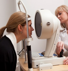
1. The preoperative exam:
First of all exact measurements are done of the refractive alteration which has to be corrected. Contact lens wearers remove their contacts around a week before the exam (soft contact lenses), and 2 to 3 weeks before the exam for hard contact lens wearers.
The parameters, which will give us the correct preoperative protocol are: the curvature of the cornea (keratometry), the thickness of the cornea (pachymetry), the diameter of the cornea, the pupil diameter, the axial length lens of the eye (echography), the eye pressure, the age and the sex of the patient and the dominance of the eye. The patient’s eyes may not have absolutely any kind of previous disease, and the central and peripheral retina has to be intact. The topography of the cornea gives us a very exact mapping of the cornea, with which a surgical protocol can be refined, and therefore the postoperative follow-up much better studied as well as eventual retreatments planned.
The day of the surgery
Procedure
- Remove your soft contact lenses 2 weeks before surgery.
- Hard or oxygen-permeable contact lenses must be removed at least 1 week in advance especially if you have been wearing them for a long time.
One day before surgery:
- Get the prescribed drops from the pharmacy.
- No make-up.
- Contact your driver.
On the day of the surgery:
- Bring with you: the prescribed drops.
- No make-up or perfume.
- The examination and the surgery will take approximately 1 hour. If there is a delay in the operation list your surgery time may be retarded.
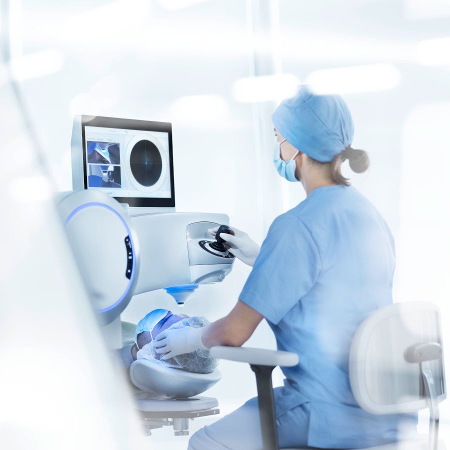
The operation:
Anesthesia: drops several minutes before.
- During surgery you will not be able to close your eyes, because a speculum keeps it open.
- During the flap making you will not see for several seconds.
- During the laser treatment an intermittent noise will be heard.
- At the end the eye is irrigated rinsed profusely.
After surgery:
- Drops will be used as prescribed during the first week.
- 4x day antibiotics.
- Natural tears hourly.
Recuperation:
- Activity will be restarted the day after surgery.
- Contrast sensitivity and reading will be difficult the first days.
- No contact sports or swimming for 3 weeks.
- No make-up for 3 weeks.
Post-operative appointments:
- 1st day
- 1 week
- 1 month
Please arrive at the time indicated

2. Treatment:
b. After having made the cornea flap with a very fine instrume
nt, which is called a microkeratome, this flap is lifted aside to give room to the laser to be performed right afterwards.
· Laser:
The excimer laser has a concentrated beam of light, which can precisely be directed to a certain point. With the help of very fine laser pulses, microscopic quantities of tissue of the cornea are ablated. With this laser property, the cornea curvature becomes the desired curvature and therefore the defect is treated. The duration of the laser application is between 1-2 minutes.
· Eye tracker:
During the laser treatment we always use an eye tracker. The computerised eye tracker directs exactly the laser beams up to the smallest eye movements to get a very exact treatment. The treatment has a direct result and is without pain, as the treatment is performed in the deep layers of the cornea. The eyesight recovers in general the same day and is normally within a week optimal.
3. Postoperative:
Immediately after the intervention, the patient stays around 20 minutes resting. Afterward the first ophthalmic control is performed. While this control is done, the patient may go home: the eyes are not covered and the eyesight reaches already around 80% of the total capacity.
It is advised after the intervention not to rub the eyes during several hours. The next day the eyesight is clearly better.
During one week after surgery eye drops are applied in the eyes.
4. Controls:
There is a very quick healing and eyesight recovery. Most of the kind the patient can recover his normal activities within 24 hours after intervention. Controls are planned the day after surgery, one week after and 3 weeks after surgery.
5. Personalized ablations:
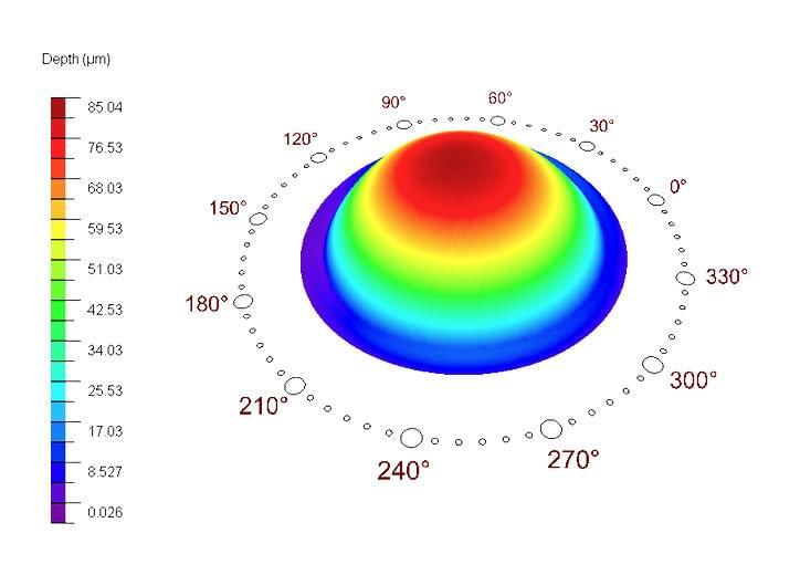
a. Topography and aberrometry
With classic corneal topography we can study the superficial part of the cornea. This gives us the study of the corneal curvature.
The newest and most exact elevation topographers like the Orbscan, Pentacam, gives us the study of the cornea more in the depth. In this way we can study the profile of the cornea from the front part and the back part of its surface and therefore to reconstruct its whole depth. At the same time we can also measure its curvature as well as a pachymetric map, which gives us the depth of the cornea in different points.
The topographical examination over the cornea gives us in a very exact way the curvature of the cornea being able to evaluate all kind of alterations to be able to correctly plan treatments and at the same time avoid contraindications in the refractive surgery.
Topographical examinations are used before and after surgery to be able to do exactly follow-ups.
Wave front technology is used by astronomers since long ago to perfectionate their telescopes. The image that comes back from the retina in a wavefront analysis is not perfectly due to the different anomalies of the surroundings such as cornea, tear film, anterior chamber, lens, aqueous humor. The image that comes back from a detector of a telescope is also distorted due to the earth atmosphere. Therefore these problems are comparable. The aberrations of the eyesight are comparable with the turbulence of the atmosphere. This technology is used in ophthalmology to be able to analyse all eye aberrations and at the same time treat them with the excimer laser beams.
Femtolasik
The newest technique in our treatment. Called also “All-laser Lasik” or “Bladeless Lasik”. Is a technique added to the Lasik treatment to create a thinner flap, when necessary, through photo disruption guided by computer software.
We use the Zeiss Visumax 800
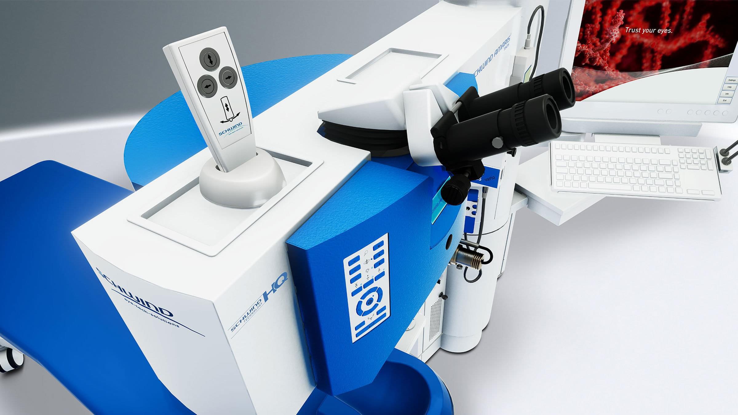
Schwind Amaris 1050 RS
To stay with the most advanced technology the Schwind Amaris 1050 RS laser excimer.
This is the most advanced laser in the market:
- The fastest currently available (1050 Hz).
- 6-dimensional turbo eye tracker (1050 Hz) that follows every movement through an infrared high speed camera.
- The treatable optical zone is 10 mm, in comparison to only 8 mm for other excimer lasers. This reduces the risk of halos and other disturbing visual phenomena after laser treatment.
- Wavefront adjusted and corneal wavefront technology is integrated as standard in the eye laser treatments. This technology assures the quality of normal vision, night vision and contrast vision. Every treatment is therefore completely customized.